
Performing right heart caths
Right heart catheterization—or right heart cath—is the only test that can confirm a pulmonary arterial hypertension (PAH) diagnosis. In contrast to right heart echoes, right heart caths provide direct measurements of key hemodynamic parameters and pressures and, when used over time, can track a patient's disease progression and treatment responses.1
Normalizing hemodynamics and improving right heart size and structure are critical to ensuring optimal long-term outcomes in your patients with PAH. Monitoring and understanding these hemodynamic parameters helps ensure optimal treatment outcomes by proactively adjusting therapies when a patient hasn’t sufficiently improved.2
Key hemodynamic parameters
Pulmonary artery pressure (PAP), pulmonary arterial wedge pressure (PAWP), and signals of right ventricular (RV) dysfunction measured by right heart cath provide critically important prognostic information about your patient’s disease status. These measurements should be done at diagnosis and periodically during follow-up over the course of treatment.1-5
Additional hemodynamic parameters that provide a comprehensive view of the right heart include:
BNP/NT-proBNP
CI
CO
mPAP
PVR
RVSP
Right heart cath is required for diagnosis and baseline data1
Right heart cath is the only test that can confirm a PAH diagnosis. When clinical tests and echo imaging indicate a suspicion of PH, right heart cath is required to confirm diagnosis. Data from the right heart cath at diagnosis help differentiate which form of PH a patient may have and help guide selection of the initial treatment regimen.1
Right heart cath is needed to confirm PAH
Many clinical indicators can increase the likelihood that your patient may have PAH, including signs and symptoms, lab tests, and a virtual echocardiography screening tool (VEST) score derived from echo imaging data. Any of these results may lead you to request a right heart cath for your patient to confirm a PAH diagnosis.6
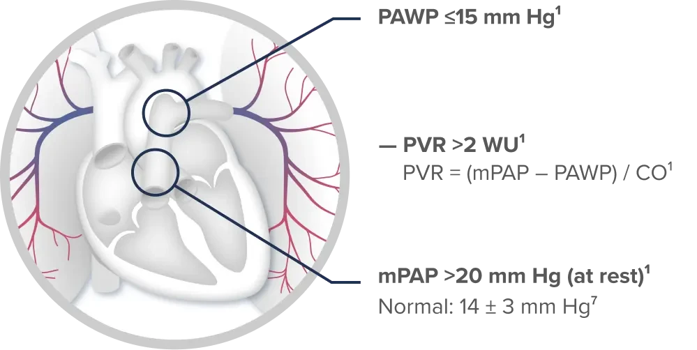
Based on today’s guidelines, confirmation of PAH is when a patient has1:
- mPAP >20 mm Hg
- PAWP ≤15 mm Hg
- PVR ≥2 WU
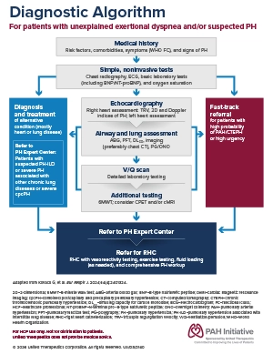
Review the diagnostic algorithm based on current guidelines and recommendations
Download Diagnostic AlgorithmBaseline right heart cath data inform treatment plans
The pressure readings and hemodynamic data gathered from right heart cath can be used to complete a formal risk calculation for patients diagnosed with PAH. Today’s treatment guidelines use the baseline risk score as the primary input to determine the combination of medications used in the initial treatment regimen.2
Right heart cath for clinical monitoring for PAH
Right heart pressures are critical in diagnosing and setting initial treatment plans and can be helpful in monitoring your patients’ right heart function once they are on therapy. The current 2022 ESC/ERS Treatment Guidelines recommend more frequent evaluations: right heart cath should be ordered in cases of clinical worsening and can be used for follow-up 3 to 6 months after changes in therapy.1
Right heart cath evaluations can show patient improvement and responses to treatments. They can also reveal any hemodynamic worsening, serving as a trigger to escalate treatment to help your patient’s long-term outcomes.1

Learn how key hemodynamic parameters help determine patient disease status and treatment response
Watch Short VideoHemodynamics from right heart cath are prognostic
The 2022 ESC/ERS Treatment Guidelines–based risk strata are among the many registries representing real-world data captured from thousands of patients across global registries and clinical practice. These data are reliable predictors of how well your patient is doing.1,3,8-10 Because hemodynamic variables provide prognostic data, they are embedded in these risk calculation tools, such as the REVEAL 2.0 or 2022 ESC/ERS calculator. Managing PAH with these validated, real-world parameters helps clinicians navigate the complexities of PAH and choose the combination treatment regimen that is appropriate for their patients’ disease status.1,2
Risk of 1-year mortality from 2022 ESC/ERS Treatment Guidelines1
Low risk
<5%
Intermediate risk
5%-20%
High risk
>20%
Normal right heart cath pressures have clinical significance
Understanding key right heart cath pressures is essential because they provide a definitive assessment of hemodynamic function and strain. These measurements allow clinicians to accurately stratify patient risk and tailor treatment decisions. When hemodynamic parameters remain elevated or worsen despite current therapy, escalating treatment by adding agents that target additional pathways should be considered to improve hemodynamic function and prevent disease progression.1
Parameters to manage PAH
Parameter
Normal values
Clinical significance
Measured parameters
RAP1,11
2-6 mm Hg
Reflects RV preload & degree of right heart venous congestion
RVSP12
15-30 mm Hg
Represents peak pressure generated by RV during systole
mPAP1,12,13
8-20 mm Hg
Represents average pressure in PA throughout cardiac cycle
sPAP1,14
15-30 mm Hg
Represents peak pressure during RV systole
dPAP1,14
4-12 mm Hg
Reflects residual pressure during diastole
PCWP14
6-12 mm Hg
Indirectly estimates LAP & LV filling pressure; differentiates precapillary from postcapillary PH
CO1,15
4-8 L/min
Reflects total volume of blood pumped per minute
SvO21,2
65%-80%
Measures overall oxygen status; in PAH, low SvO2 is associated with declining RV function
Calculated parameters
CI1
2.5-4.0 L/min·m2
Reflects right heart function; CI = CO / BSA
PVR1,2
≤2 WU
Reflects RV afterload & burden on RV function
Get the Right Heart Cath Quick Reference
Download Right Heart Cath ParametersRight heart cath: A routine procedure critical to patient management
You can help patients understand the importance of securing accurate information to establish a proper diagnosis and treatment plan.
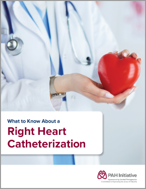
The PAH Initiative has developed a patient education brochure to help you explain the RH cath procedure. This information may help you align expectations and provide patients with a clear understanding of what to expect during the procedure.
Where can my patients get a right heart cath?
If you do not have a relationship with clinicians who conduct right heart caths or are looking for right heart cath facilities, the CHEST PH Expert Center Finder is a helpful resource to explore. Below is a link that helps you locate providers near you who can perform right heart caths. CHEST is a global leader in advancing best patient outcomes through innovative chest medicine education, clinical research, and team-based care.
Connect With a PH Expert in Your Area