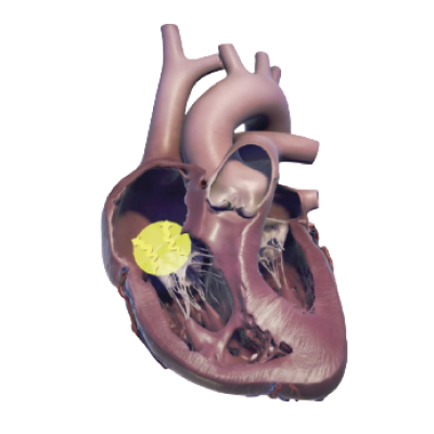
Echoes for Pulmonary Hypertension

Echo findings reveal early changes in the right heart

The advances in transthoracic echo technology (also called TTE technology), the practicality of this noninvasive tool, and the critical reproducibility of measuring size, structure, and function of the right heart demonstrate the crucial value of echo imaging in managing pulmonary arterial hypertension (PAH).1

Noninvasive echo assessments of the right heart are a critical component of patient management and have 2 key roles: input into the diagnostic process and ongoing, frequent monitoring of disease progression and prognosis.1

PAH likelihood

Readily accessible right heart echoes are often the first view into the likelihood of PAH.1

Combined with clinical signs, adding echoes as part of the diagnostic workup helps identify abnormal parameters that signal the need for right heart cath (right heart catheterization) to make a PAH diagnosis.1,2

PAH monitoring

Frequent echo monitoring of the right heart gives clinicians critical knowledge to monitor disease status and make timely treatment decisions.1

Right heart echoes also provide key inputs to help risk-stratify patients and understand their prognosis, ensuring right heart health is factored into treatment plans.1

Right heart echo is a critical prognostic and monitoring tool

Though PAH is a progressive lung disease, the changes in the pulmonary vasculature directly affect the right side of the heart, particularly the right ventricle.1,2 The resulting abnormal modifications in right heart size, structure, and function are indicative of your patient’s overall disease status.1

Frequent and comprehensive TTE monitoring is like an early-warning system—detecting changes in the right heart before clinical symptoms appear or as the heart moves to near-normalization.3-5 Accurate, precise echo values help identify the subtle initial signs of abnormal right heart size, structure, and function. Catching early changes in the right heart helps clinicians make timely treatment adjustments to improve patient outcomes.1,2,4

The echo doesn’t lie

Quick Clips!

Watch as Dr. Anjali Vaidya explains the role of echo in detecting changes in right heart function.

View the video:
Echo for Monitoring PAH (11 min)

[Video title: The Echo in PAH: “The Echo Doesn’t Lie”]

[On-screen text: John Ryan, MD, MB, BCH, BAO; Anjali Vaidya, MD, FACC, FASE, FACP; Excerpt from The Echo in PAH: State of the Art in Right Heart Monitoring]

[On-screen text: Anjali Vaidya, MD, FACC, FASE, FACP, Professor, Medicine, Lewis Katz School of Medicine at Temple University; Co-Director, Pulmonary Hypertension, Right Heart Failure & CTEPH Program, Temple University Hospital, Philadelphia, Pennsylvania]

Dr. Anjali Vaidya: There is so much variability and error in the RVSP estimation for a variety of Echo Doppler technique issues, and this has been shown time and time again, particularly some data that show up to 50% of patients will have a 10 millimeter of mercury error in the Echo Doppler estimation of pressure relative to the directly measured hemodynamics. So, the bottom line is the pressure is never the answer. It's never the answer in the diagnosis. It's never the answer in the risk assessment, even less so. It's meant to raise awareness to look closer. And once you have a suspicion of it, we have to look at all of the right heart parameters, right ventricular size, structure, function, tricuspid regurgitation, right atrial size and shape, IVC collapse, and all of those things. And so, if we do our Echos more frequently, then our clinical gestalt tends to be more in line with a formal validated objective risk assessment. And I think that point is really key. So, I always tell my fellows and my residents, "The Echo doesn't lie", whereas you just nicely presented a case where 6-minute walk and Functional Class could represent such a wide range of actual cardiac performance. So it's a long-winded way of saying more frequent is better. Any sign of a clinical change in a relatively urgent Echo, if I'm escalating therapy, or if I've recently changed their therapy, then I'm not waiting more than 3 months for my next echocardiogram.

[On-screen text: Excerpt from The Echo in PAH: State of the Art in Right Heart Monitoring; View the entire program at www.RightHeartInPAH.com]

[On-screen text: PAH Initiative; Sponsored by United Therapeutics; PAHinitiative.com/hcp; © 2023 United Therapeutics Corporation. All rights reserved. US-DS-1109]


Right ventricular health is a key determinant of survival in PAH

Across multiple patient registries and thousands of patients’ data, right heart echo results have shown to be prognostic for PAH patients.1 These prognostic values offer critical insights into your patient’s disease status and allow for timely treatment decisions.

Taken together, many echo parameters, including, but not limited to, TAPSE/PASP, PVR, RAP, degree of TR, and IVC, can help evaluate and project how a patient’s disease may progress.1,2,6 Armed with this prognostic information, providers can make informed decisions about which treatments to use and how to further manage their patients to facilitate their long-term outcomes.

Data validate use of echoes in patient management

TAPSE values are highly sensitive indicators of right ventricular performance, help track right heart changes over time, and can predict prognosis.7

Video icon

Watch experts discuss how they use echoes in their clinical practice

Watch Short Video

Survival stratified by follow-up TAPSE7

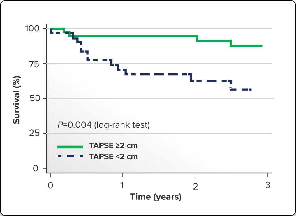
One prospective study followed 70 patients with PAH over the course of 3 years. Patients with TAPSE ≥2 cm (n=37) measured 6 months after their baseline echoes were more likely to:

  • Achieve FC I or II status
  • Demonstrate a higher 6MWD
  • Experience improved overall survival
Kaplan-Meier estimates of survival stratified by TAPSE values: better survival when TAPSE is ≥2 cm
Image Description

Kaplan-Meier estimates of survival are stratified by TAPSE values. Survival is stratified by follow-up TAPSE value <2 cm or ≥2 cm and reveals better survival when TAPSE is ≥2 cm.7

Mazurek JA, Vaidya A, Mathai SC, Roberts JD, Forfia PR. Follow-up tricuspid annular plane systolic excursion predicts survival in pulmonary arterial hypertension. Pulm Circ. 2017;7(2):361-371.

Survival stratified by echo parameters6

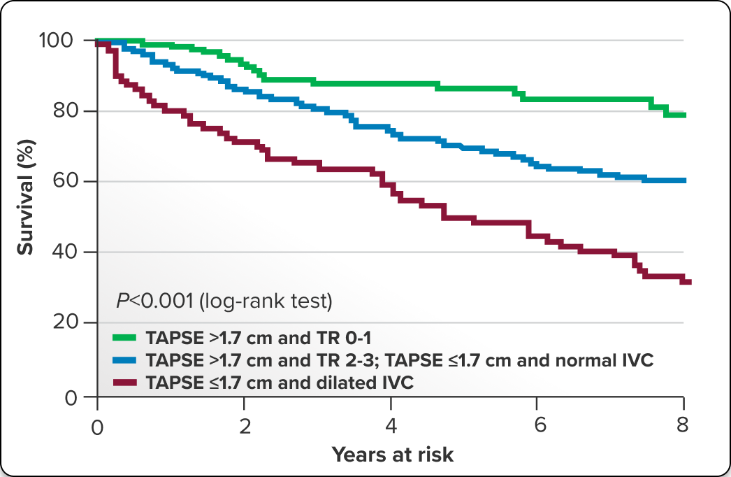
Integrating multiple parameters, such as TAPSE (>1.70 cm), degree of TR, and IVC dilation, has been valuable when stratifying the risk of mortality in patients with PAH. In a pooled analysis of 517 individual data points from 7 observational studies, patients with reduced TAPSE, reduced TR, and absence of dilated IVC were shown to have significantly better prognosis and higher survival rates.

Kaplan-Meier survival curves according to 3 echocardiographic-based risk groups in patients with PAH. Kaplan-Meier curves show significantly lower 5-year survival rates for high-risk patients (TAPSE ≤1.7 cm and dilated IVC) compared with both intermediate-risk patients (TAPSE >1.7 cm and TR 2-3; TAPSE ≤1.7 cm and normal IVC) (43% vs 63%, respectively; P<0.001) and low-risk patients (TAPSE >1.7 cm and TR 0-1)
Image Description

Kaplan-Meier survival curves according to 3 echocardiographic-based risk groups in patients with PAH. Kaplan-Meier curves show significantly lower 5-year survival rates for high-risk patients (TAPSE ≤1.7 cm and dilated IVC) compared with both intermediate-risk patients (TAPSE >1.7 cm and TR 2-3; TAPSE ≤1.7 cm and normal IVC) (43% vs 63%, respectively; P<0.001) and low-risk patients (TAPSE >1.7 cm and TR 0-1) (43% vs 82%, respectively; P<0.001).6

Ghio S, Mercurio V, Fortuni F, et al; TAPSE in PAH Investigators. A comprehensive echocardiographic method for risk stratification in pulmonary arterial hypertension. Eur Respir J. 2020;56(3):2000513.

Echoes reveal PAH prognosis

Evidence across multiple registries and clinical studies demonstrates that right heart echo parameters provide critical prognostic value in PAH.1,6,7 Understanding your patient’s prognosis helps assess whether sufficient improvement has been made toward the goal of near-normalization of the right heart.1

When right heart parameters indicate suboptimal response, clinical decisions and timely treatment adjustments can be initiated.8 Some key right heart parameters that have useful prognostic values are listed below.

Right heart parameter prognostic values in PAH Two-column comparison of right heart parameters and their associated prognostic values.

Right heart parameters

Prognostic values

RV fractional area change9
  • Associated with survival
  • Abnormal if ≤32%
RA area and area indexed to body surface area1
  • Predict morbidity––each 1-cm2 increase correlates with a 6% increased morbidity risk
  • RA area >26 cm2 has increased mortality risk
Pericardial effusion1
  • Marker of poor prognosis
TAPSE/PASP ratio1,10
  • <0.19 mm/mm Hg indicates a 1-year mortality risk of >20%
  • <0.22 cm/mm Hg is associated with a 37% higher mortality risk
RA pressure1
  • ≥5 mm Hg indicates poor prognosis

Key echo assessments: Size, structure, function

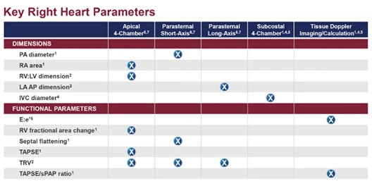
Echoes provide an objective, real-time view of right heart size, structure, and function.1 Review these descriptions of key echo assessments below to ensure that your echo results include these essential assessments:

  • Right heart size and structure
  • Right heart function
Video icon

Examine right heart echo parameters: size structure, function

Watch Short Video

Assessing right heart size and structure

When assessing right heart size and structure, an important evaluation is whether the right ventricle and/or right atrium is enlarged.1 Additionally, examining the septum in systole and assessing for signs of pericardial effusion can reveal whether a patient may have severe PAH.1 Examine the following table for details of right heart size and structure parameters.

Right heart size and structure assessments Three-column comparison of assessments, their significance, and key highlights.

Assessments

Significance

Key highlights

RV1,11
  • Provides insights into PH mechanism and how much a patient is compensated
  • Important to evaluate at least 1 measure of RV chamber size (RV:LV ratio, RV wall thickness, RV base to apex systolic ratio)
  • RV base to apex ratio <1.5 is strongly associated with elevated PVR
RA1
  • Abnormal RA dilatation reflects chronic elevated right-sided heart pressures
  • RAP is measured looking at the collapse of the IVC rather than looking at the RA
Septal flattening in systole1
  • Evaluates presence and extent of RV volume
  • Represents elevated afterload to the RV (ie, PVR)
  • Even mild systolic septal flattening is abnormal
  • May be among the first signs of PAH
PA diameter1
  • Main PA dilates in response to pressure and volume overload
  • Abnormal diameter >25 mm may indicate increased RV afterload
  • PA diameter:ascending aorta diameter ratio ≥1 is abnormal
RV:LV dimension1
  • Comparison of RV to LV size
  • Normal RV is <2/3 size of LV
  • RV:LV ratio >1 (basal) suggests moderate RV dilation
LA volume index1,12,13
  • Simple-to-measure indicator of LA size
  • Normal LA volume can help distinguish precapillary from postcapillary PH

Review your own right heart echo protocols to ensure you receive the critical information needed to assess and manage your patients with PAH

Assessing right heart function

When assessing right heart function, focus on 3 primary measures: TAPSE, RVOT, and TRV.

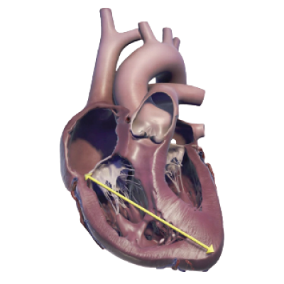
TAPSE diagram

TAPSE (tricuspid annular plane systolic excursion)1

Tap or click to learn more

Easiest way to assess the overall RV’s longitudinal movement during systole (contraction)

  • Normal threshold >1.70 cm
    • Below-normal threshold indicates RV dysfunction and poor prognosis
  • With PASP, measurements represent the degree of RV:PA coupling
  • Normal TAPSE:PASP ratio is 0.5-0.7 mm/mm Hg; if lower, there may be uncoupling
Tap or click to flip back
RVOT diagram

RVOT (right ventricular outflow tract)1

Tap or click to learn more

Doppler shows the reflected wave that occurs before the pulmonic valve closes at the end of systole

  • If there is shortened acceleration time, look for changes in the waveform, such as mid-systolic notching, shortened time to peak contraction, and a reduced acceleration time
    • These are associated with elevated PVR and increased afterload
Tap or click to flip back
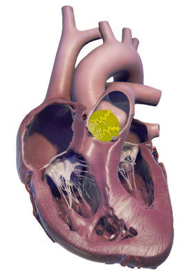
Peak TRV diagram

Peak TRV (tricuspid regurgitation velocity)1,6

Tap or click to learn more

Used to estimate PASP and RAP

  • An important measurement when investigating the likelihood of PH
    • TRV ≥2.8 m/s suggests PH likelihood, but only when validated by at least 2 other abnormal echo signs
  • Degree of TR can also be used in risk-stratifying previously diagnosed patients when combined with TAPSE and degree of IVC dilation
Tap or click to flip back

Estimating RVSP from echo

Echo RVSP is an estimation of the RV pressure, calculated from TRV measurements using a modified Bernoulli principle. Although estimated RVSP can be helpful when initially screening for likelihood of PH, it is a less relevant parameter for consideration after diagnosis. It is not a key input when determining disease status after diagnosis, nor is it a reliable factor when making treatment adjustment decisions. Other, more direct measurements of right heart size, structure, and function have become standard for long-term, ongoing monitoring.1

  • In estimated RVSP derived from echo measures, even small errors from the best labs can be amplified, leading to both under- and overestimation of actual pressures1

Focus on direct measures of right heart size, structure, and function to determine/inform PAH disease status

What are normal values?

Understanding normal echo values is critical to knowing when it is time to look deeper into your patient’s right heart parameters. For instance, knowing that a normal RV:LV ratio is <0.7, a normal TAPSE is >1.70 cm, and a normal RVSP is ≤34 mm Hg can aid in evaluating your patients. When echo values are not normal, further investigation is required.1

If your patient’s echo values are not normal, that should increase your concern that your patient has not achieved near-normalization of the right heart. It may be that the treatment plan is not sufficiently improving the patient’s disease status and treatment escalation should be considered.2,8

Normal right heart echo assessment values1

Right heart parameters
Normal values
RV
  • RV:LV ratio <0.7
  • RV wall thickness <5 cm
RA
  • RA size and area <19 cm2
TAPSE
  • TAPSE >1.70 cm
  • TAPSE/PASP ratio 0.5-0.7 mm/mm Hg
RVOT
  • RVOT VTI >18 cm
Peak TRV
  • ≤2.8 m/s
RVSP
  • ≤34 mm Hg

With a goal of achieving low-risk status in the first year of therapy, learn about treatments that may improve right heart parameters2,14

Download key right heart echo parameters

Download Parameters

Today’s treatment guidelines recommend frequent echo monitoring

Due to mounting data over the years, it is clear that right heart size, structure, and function crucially inform PAH disease status and can be an early indication of disease progression.1,3,4 Recent advances in understanding right heart hemodynamics inform the newest standard of care for patients with PAH, which includes frequent monitoring using echoes every 3 to 6 months. This ongoing assessment is vital for patients who have changed treatments or experienced clinical worsening or who even appear stable.1,2,4

Current guidelines, including both the 2022 ESC/ERS Treatment Guidelines and the 2024 WSPH Recommendations, now incorporate this standard of care for right heart echoes.2,8

Photograph of Anjali Vaidya

“[Echo] monitoring is really critical [in PAH], and it should be done with high frequency to identify early recognition of a patient’s clinical status that’s declining.”

— Anjali Vaidya, MD, FACC, FASE, FACP, Cardiologist

Useful resources for comprehensive right heart echoes

The PAH Initiative wants to support you in gaining access to accurate and complete right heart echoes.

The American Society of Echocardiography (ASE) is a great resource to dive into the technology and techniques of echoes and offers educational tools to enhance your understanding of echoes.

Additionally, the ASE publishes standards for creating consistent, comprehensive echo reports that clearly inform clinicians on the health status of a patient’s heart.

Echoes have a critical role in evaluating patients with PAH

This content was developed based on ASE Guidelines.

6MWD=6-minute walk distance; echo=echocardiography; ERS=European Respiratory Society; ESC=European Society of Cardiology; FC=functional class; IVC=inferior vena cava; LA=left atrium/left atrial; LV=left ventricle/left ventricular; PA=pulmonary artery/pulmonary arterial; PASP=pulmonary artery systolic pressure; PH=pulmonary hypertension; PVR=pulmonary vascular resistance; RA=right atrium/right atrial; RAP=right atrial pressure; RV=right ventricle/right ventricular; RVSP=right ventricular systolic pressure; TR=tricuspid regurgitation; VTI=velocity time integral; WSPH=World Symposium of Pulmonary Hypertension.
References: 1. Mukherjee M, et al. J Am Soc Echocardiogr. 2025;38(3):141-186. 2. Humbert M, et al. Eur Heart J. 2022;43(38):3618-3731. 3. Adamson PB. Curr Heart Fail Rep. 2009;6(4):287-292. 4. Milks MW, et al. J Heart Lung Transplant. 2021;40(3):172-182. 5. Simonneau G, et al. Am J Respir Crit Care Med. 2002;165(6):800-804. 6. Ghio S, et al; TAPSE in PAH Investigators. Eur Respir J. 2020;56(3):2000513. 7. Mazurek JA, et al. Pulm Circ. 2017;7(2):361-371. 8. Chin KM, et al. Eur Respir J. 2024;64(4):2401325. 9. Howard LS, et al. Eur Respir Rev. 2012;21(125):239-248. 10. Mukherjee M, et al; PVDOMICS Study Group. Circ Cardiovasc Imaging. 2024;17(10):e017074. 11. Labrada L, et al. Curr Opin Pulm Med. 2023;29(5):348-354. 12. Opotowsky AR, et al. Circ Cardiovasc Imaging. 2012;5(6):765-775. 13. Vaidya A, et al. Pulm Circ. 2020;10(3):1-10. 14. Boucly A, et al. Eur Respir J. 2017;50(2):1700889.

Going to CHEST?

Can first-year treatment decisions really change PAH outcomes?

Hear experts explore the data

The Clinical Challenge: How Important Is the First Year of PAH Therapy?

Monday, October 19, 2026

2:30 ᴘᴍ–3:15 ᴘᴍ

Phoenix Convention Center

Learning Theater 3

Hear 4 PAH experts explore key considerations in patient care today:

  • Why low risk matters
  • How early action can help improve outcomes
  • How frequent risk assessments and right heart evaluations can inform treatment decisions