
Conduct comprehensive right heart echo assessments
The ASE Guidelines issued the Echocardiographic Assessment of the Right Heart (PDF) in March 2025. These guidelines reflect advances in pulmonary arterial hypertension (PAH) care and the critical role right heart assessment plays in patient management. The guidelines also provide details on the updated PH classification definitions, descriptions of the optimal acoustic windows, echo views, and measured parameters that are critical for a comprehensive right heart evaluation. Collecting these crucial right heart echo images and values is paramount in helping define PAH disease status and determine treatment plans.1
Today’s echo assessments go far beyond estimating pressures like RVSP; instead, direct, reliable echo measurements of the right heart size, structure, and function are key to recognizing your patient’s disease status and prognosis.1 Understanding right heart parameters is crucial to making patient management decisions, as these parameters can predict patient survival.2,3
Additionally, ASE Guidelines emphasize the importance of frequent, systematic assessment of right heart size, structure, and function. Serial monitoring—typically every 3 to 6 months—provides clinicians with key parameters to more thoroughly assess PAH disease status and changes in patient progression as they respond to treatment or to know whether patients need treatment adjustments.1
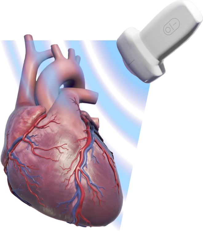
Clinicians depend on accurate right heart echoes
Right heart echoes are used in 2 critical areas in PAH1:
- When evaluating the probability of PAH
- When performing frequent, serial assessments during PAH management

Watch how echoes are used to evaluate the likelihood of PAH
Watch Short VideoHoning the probability of PAH
Right heart echo imaging is critical in evaluating the likelihood of PAH. When a clinician is suspicious of PH or notes “shortness of breath” on an echo order, patients need a complete transthoracic echo evaluation, including a comprehensive assessment of the right heart. Because early symptoms of PAH overlap with left heart disease symptoms, it is critical to investigate key right heart parameters, including right heart size, structure, and function.4
Echo alone is not sufficient for definitive confirmation of PAH, as right heart catheterization (right heart cath) remains the gold standard required for diagnosis. To help determine the need for right heart cath, clinicians may use the virtual echocardiography screening tool (VEST), which is derived from echo results. The more “yes” results, the higher the probability a patient may have PH.5
Echo parameter5
Yes
No
Mitral E:e′, lateral ≤10 (or E ≤90 cm/s)
+1
–1
Qualitative left atrial size normal or mildly enlarged
+1
–1
Systolic interventricular septal flattening
+1
–1
Frequent, serial echoes for PAH management
Clinical management of PAH relies on a comprehensive measurement of right heart parameters.1,4
When performing an echo on a patient with PAH, the focus must be on a comprehensive assessment of the right heart to determine its size, structure, and function—critical parameters that are integral to clinicians when managing PAH patient care.1,4
This page summarizes the ASE Right Heart Echocardiography Guidelines, with key tomography and right heart imaging processes and parameters that a PAH clinician should order for their patient with PAH.
4 key views for optimal right heart echo imaging
Only 4 key tomographic views are required to provide the information clinicians need to assess the right heart.6 Since right heart echoes may not be as routine as left heart echoes, the following overview and probe placement tips help optimize right heart echo imaging views to convey the best echo results for clinicians.
Tomographic views for right heart echoes

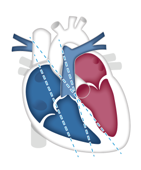
Apical 4-chamber imaging6-8
Structures
- Left ventricle (inferior septum and anterior lateral segments)
- Right ventricle
- Left atrium
- Right atrium
- Mitral valve
Probe placement
Ensure the probe is placed at the cardiac apex, with the marker angled toward the patient’s left side
Parasternal long-axis imaging6-8
Structures
- Right atrium
- Tricuspid valve
- Pulmonary valve
- Pulmonary artery
Probe placement
Ensure the probe is placed at the left sternal border, with the marker angled toward the patient’s right shoulder
Clinicians may also request views of the right ventricular inflow and outflow tracts
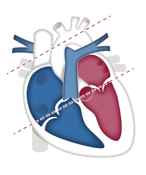
Parasternal short-axis imaging6-8
Structures
- Pulmonary valve
- Pulmonary artery
- Mitral valve
- Interventricular septum
- Right ventricle
Probe placement
Ensure the probe is placed at the left sternal border, with the marker angled toward the patient’s left shoulder
Clinicians may also request views at the aortic valve and mid-cavity levels

Subcostal 4-chamber imaging6-8
Structures
- Right atrium
- Right ventricle
- Tricuspid valve
- Pulmonary valve
Probe placement
Ensure the probe is placed below the xiphoid process, with the marker angled toward the patient’s left side
View key tomographic views for echo imaging and measurements in this animated video
Watch Short Video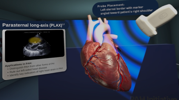
Key right heart echo size and structural measurements
These cards review the 2025 ASE Guideline recommendations of the critical right heart images and measurements that should be included in a standard right heart echo report.
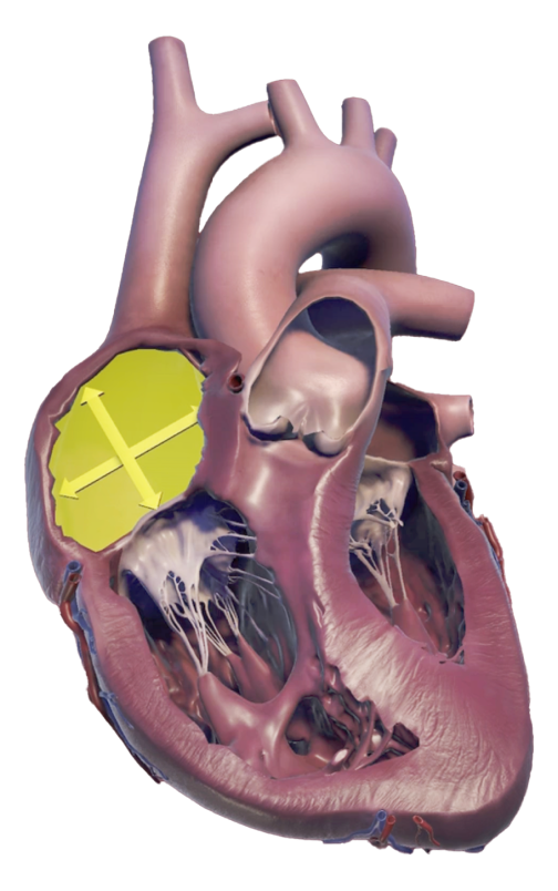
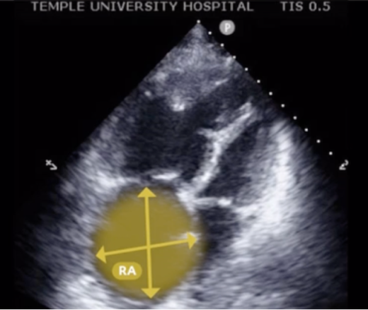
Right atrium1
Imaging: The right atrium can be optimally imaged from the apical 4-chamber view at the end of ventricular systole and tilting upward, maximizing the superior aspect of the chamber.
RA volume assessment is best using the single-plane method of discs approach.
Reporting: Include the RA area and right atrial volume indexed to body surface area.
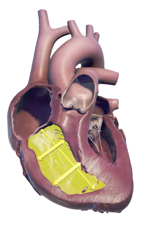
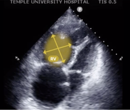
Right ventricle1
Imaging: Quantify the RV chamber using the parasternal windows and apical RV-focused view, allowing the RV free wall to be seen throughout the entire cardiac cycle.
RV wall thickness is imaged by subcostal view through direct 2DE measures and/or M-mode or by PLAX view.
Reporting: Include at least 1 quantitative measure of RV chamber size, although multiple measurements may be needed. Include RV wall thickness in the report if it is abnormal (≥5 mm).
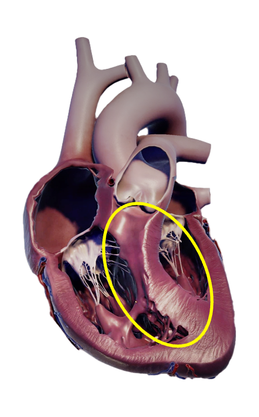
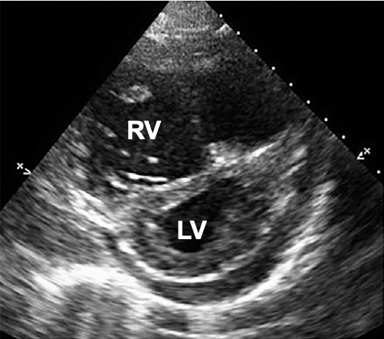
Interventricular septum1
Imaging: Prognostic in PH, the LVEI is the ratio of LV anteroposterior and LV septolateral values measured using PSAX view at the papillary muscles.
Reporting: LVEI should be reported if abnormal (>1).
LVEI >1 at end-diastole suggests RV volume overload, whereas if at end-systole, it suggests RV pressure overload. If LVEI >1 at both end-diastole and end-systole, RV pressure overload may occur with or without volume overload.
If there is septal flattening in systole, it is a sign that the RV is under severe pressure overload.
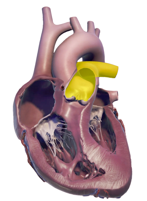
Pulmonary artery1
Imaging: MPA is measured using 2DE.
MPA diameter is measured using PSAX view, with PA-focused view in end-diastole, halfway between pulmonary veins and bifurcation of the MPA.
Reporting: MPA diameter should be reported when abnormal: end-diastolic diameter of >25 mm or MPA:ascending aortic diameter ratio ≥1.
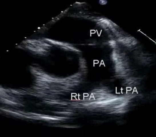
Image from Echocardiographer.org.
Evaluate normal and abnormal echo parameters critical for PAH assessments and prognosis in this video
Watch Short Video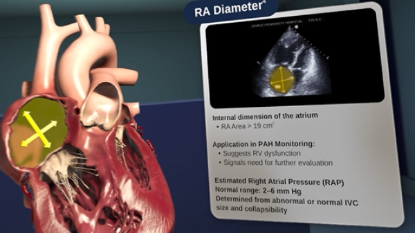
Key right heart echo functional criteria
When assessing right heart function, focus on 3 essential measures: TAPSE, RVOT, and TRV.
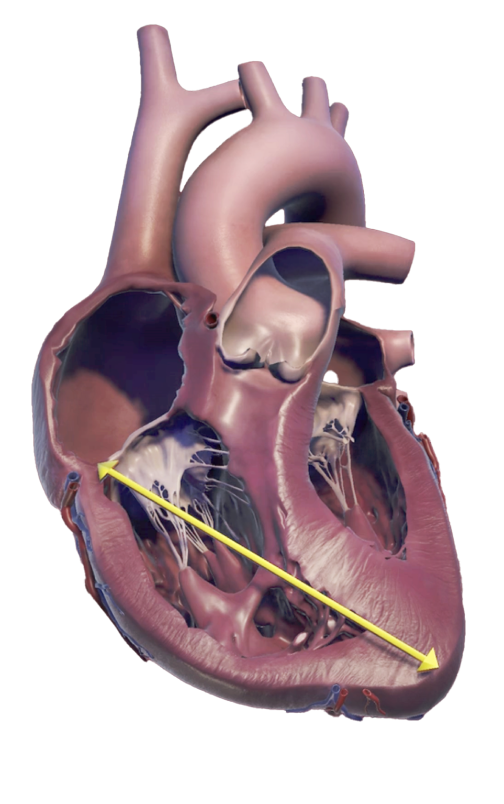
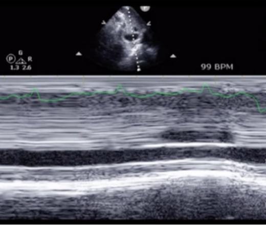
TAPSE1
Easiest way to assess the RV’s longitudinal movement during systole.
Imaging: Using the RV-focused view, image TAPSE by putting the M-mode cursor along the lateral tricuspid valve annulus.
Reporting: As a core measure of the RV, include TAPSE values within the context of RV functional assessment.
Combine TAPSE with PASP, as TAPSE/PASP indicates the RV contractile response to pulmonary afterload.
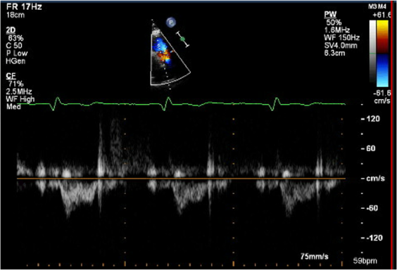
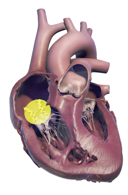
RVOT1
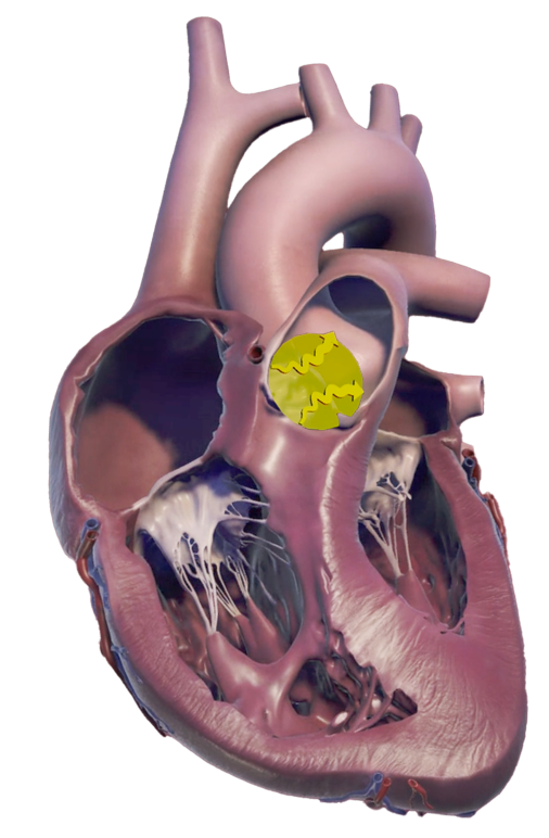
Provides valuable information on how pulmonary pressure affects the flow into the pulmonary artery.
Imaging: When imaging RVOT using Doppler, ensure the cursor is parallel to the blood flow.
Reporting: Look for specific changes in the RVOT waveform, such as mid-systolic notching and a reduced acceleration time, which are signs of increased pulmonary afterload.
TRV1
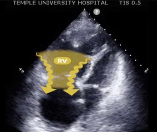
While TRV is used to understand the likelihood of PAH, the degree of TR can reveal the risk for all-cause mortality when used with other echo measurements.
Imaging: Use a continuous-wave Doppler to measure the TRV.
Reporting: Peak TRV is used to estimate other hemodynamic pressures, including RAP, PASP, and RVSP. Peak TRV ≥2.8 m/s increases the suspicion of PH, with the addition of 2 other abnormal echo signs.
A note about echo-derived RVSP
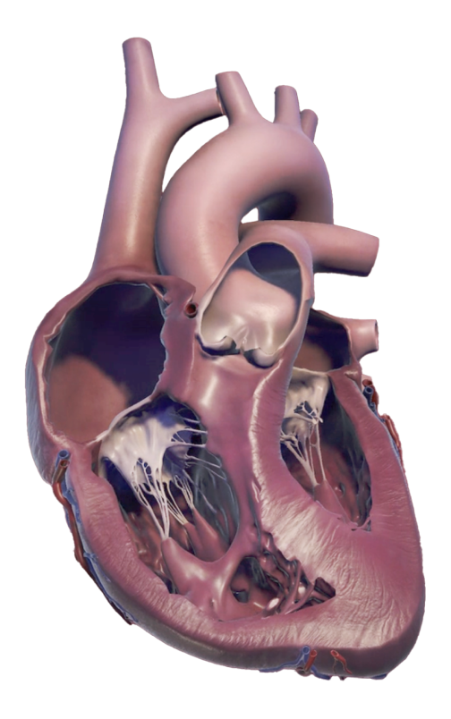
Though clinical practice has historically relied on estimating RVSP from echoes to determine a normal RVSP, today’s ASE Guidelines focus more on evaluating direct measurements of size, structure, and function as clinically reliable parameters. This shift is a result of extensive data demonstrating patient outcome measures, including survival, based on these direct measures. Estimating RVSP from echo can still play a role when initially evaluating for the likelihood of PAH but is less useful due to the wide variability in the results and inherent limitations of the estimation.1
Estimated RVSP
Imaging1: The RVSP is estimated from peak TRV using the modified Bernoulli principle, based on the following calculation:
4 × (peak TRV)²cosθ + RAP
Note that small errors may be easily exaggerated if the cursor is not parallel to flow.
Reporting1,4: Estimated RVSP is used to help determine the likelihood of PAH, but RVSP calculations may lead to under- and overestimation of actual pressures. An abnormal RVSP may be a signal for clinicians to explore potential PH and consider referring patients for right heart caths.

Resources
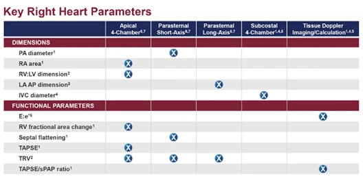
Right heart parameters
Use this guide to review key right heart parameters relevant to PAH management.
Download Echo Parameters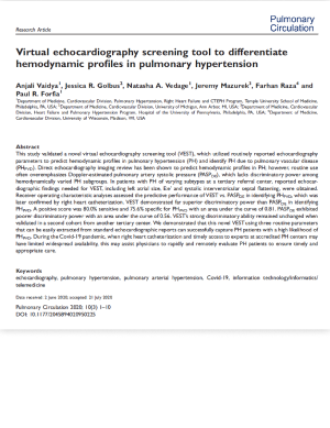
VEST score
Investigate the virtual echocardiography screening tool showing the likelihood of PAH.
Download VEST PaperRight heart caths are critical for diagnosis and monitoring
Explore the Right Heart CathThis content was developed based on ASE Guidelines.