
Near-normalization is a key treatment goal1,2
Near-normalization of the right heart—which reflects both anatomic and functional improvements of the heart—is associated with improved risk status and is critical for preventing right-sided heart failure in patients with pulmonary arterial hypertension (PAH).1,3,4 Normalizing right heart size and improving right heart function, including PVR and RVSP, are important treatment objectives to ensure optimal outcomes in your patients with PAH.1,5-7

View experts using right heart parameters to inform treatment goals
Watch Short VideoWhy is near-normalization important?
Quick Clips!
Watch as Dr. Anjali Vaidya and Dr. John Ryan describe the importance of near-normalization of the right heart as a treatment goal in PAH.
View the video:
Right Heart Imaging in Clinical Practice (10 min)
[Video title: The Echo in PAH: Near-Normalization Improves Prognosis]
[On-screen text: John Ryan, MD, MB, BCH, BAO; Anjali Vaidya, MD, FACC, FASE, FACP; Excerpted from The Echo in PAH: State of the Art in Right Heart Monitoring]
[On-screen text: Anjali Vaidya, MD, FACC, FASE, FACP, Professor, Medicine, Lewis Katz School of Medicine at Temple University; Co-Director, Pulmonary Hypertension, Right Heart Failure & CTEPH Program, Temple University Hospital, Philadelphia, Pennsylvania]
Dr. Anjali Vaidya: We want to frequently monitor this and supplement it with our kind of integrated Echo Doppler assessment to help inform our treatment decisions.
It might guide us to be more proactive and progressive in our treatment escalation, than if we solely relied on Functional Class and 6-minute walk. We know that these changes to the right heart can often occur before the subjective changes, or the functional capacity may definitively change, and we certainly see that time and time again in our clinical practice. And it's really important to note that while patient symptoms are so important for them and for us, if we wait for them to worsen before we adjust their treatment regimen, we may miss an opportunity to improve their functional impairment.
[On-screen text: Excerpted from The Echo in PAH: State of the Art in Right Heart Monitoring; View the entire program at www.RightHeartInPAH.com]
[On-screen text: PAH Initiative; Sponsored by United Therapeutics; PAHinitiative.com/hcp; © 2023 United Therapeutics Corporation. All rights reserved. US-DS-1107]
Study demonstrates extended survival when hemodynamics improved
Boucly and colleagues examined how having good vs poor hemodynamic measurements, stratified by SVI and SvO2 values, affected the transplant-free survival of 393 patients who were at intermediate-low risk at first reevaluation. They found that8:
- 286 patients with good hemodynamic function had a higher 1- and 3-year transplant-free survival of 97% and 81% (P=0.002)
- 107 patients with worse hemodynamic function had a lower 1- and 3-year transplant-free survival of 94% and 69%
These data underscore the importance of near-normalization of the right heart, showing that improved hemodynamic function resulted in better survival for these patients who were at intermediate-low risk.8
Survival outcomes stratified by hemodynamics8
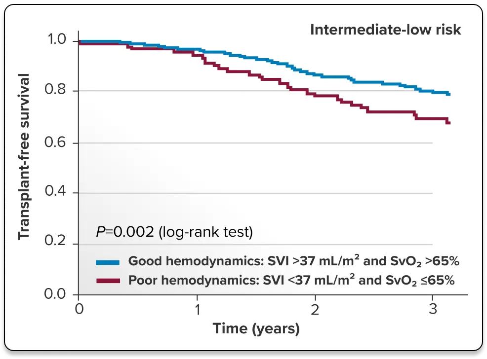
Kaplan-Meier survival curves at first follow-up risk status according to hemodynamic variables in 1019 PAH patients with at least SVI or SvO2 available at first follow-up reassessment for intermediate-low-risk status.8
A good hemodynamic profile was defined by ≥1 criterion among SVI >37 mL/m2 and SvO2 >65%. A poor hemodynamic profile was defined as neither SVI >37 mL/m2 nor SvO2 >65%.8
Measuring right heart performance is essential to PAH management
Assessing right heart performance is a foundational component in the ongoing management and risk stratification of patients with PAH. Effective PAH management requires a multiparameter approach that moves beyond physical symptoms.9 Integrating the gold-standard precision of right heart catheterization (RHC; also called right heart cath), supplemented with frequent echo monitoring plus the sensitive biochemical signaling of natriuretic peptides (BNP/NT-proBNP), reveals a comprehensive view of a patient’s disease status.1
Multimodal parameters for PAH disease

Hemodynamic performance via right heart cath
- RHC remains the gold standard for initial assessment, providing precise measurements required to establish a definitive diagnosis of PH and evaluation of severity1
- RHC provides precise and direct measurements of right heart structure and hemodynamic pressures that can be reliably tracked over time (eg, in the right atrium, right ventricle, pulmonary artery, pulmonary capillary wedge)1

Size, structure, and function via echoes
- Frequently monitoring structural changes in the right heart is an important component in long-term management, as RV remodeling often provides the earliest objective evidence of disease progression7
- Longitudinal monitoring of PAH using frequent echoes every 3 to 6 months helps track the detection of structural remodeling and RV dilation before clinical symptoms appear1,2,5-7,9
- Focus on a comprehensive echo assessment of RV size, structure, and function—core elements to monitor over time1,2,5-7,9

Right heart strain via BNP/NT-proBNP
- Monitoring BNP or NT-proBNP levels provides a biochemical early-warning system for right heart strain. Elevated BNP is a clinical signal for reassessment, using echoes or RHC (with or as part of) a risk calculation, to guide timely treatment escalation before functional decline occurs1,7,10
- Maintaining low levels of, or a significant reduction in, these biomarkers is associated with improved functional capacity and long-term survival1
What is a normal RVSP?
RVSP represents an estimate of the peak pressure generated by the right ventricle during systole. While estimated RVSP is a primary metric in echo screening, it is most clinically significant when evaluated within the broader context of right heart structure and function.7,11
RVSP approximates PASP
RVSP is a stand-in for PASP, which must be measured using an RHC. RVSP is estimated by collecting TR jet velocity during an echo. It should be noted that normal RVSP varies by age.7,12
Patients who have an abnormal TR jet velocity or abnormal estimated RVSP, in addition to other echo signs of right heart strain, should be considered for RHC.1,7,13
Patient age (years)12
Normal RVSP (mean)12
Use caution with estimated RVSP
While achieving a normal RVSP is a clinical target, clinicians should use caution when interpreting echo-derived estimates of RVSP. Much variability and error can be introduced into the RVSP formula from a variety of echo Doppler technique issues.7,11 Therefore, the results may be easily over- or underestimated.7 Estimated RVSP is meant to raise awareness of PAH and encourage clinicians to more carefully evaluate their patients. Once a patient is diagnosed with PAH, ASE Guidelines recommend investigating a range of right heart parameters—size, structure, and function, to name a few—for a comprehensive understanding of your patient’s right heart health.7,14
Today’s guidelines recommend frequent right heart echo monitoring7
Using right heart echoes to evaluate right heart size, structure, and function allows for early identification of abnormal right heart measurements that signal a need for therapy adjustment even before clinical changes are evident.1,2,9 The 2022 ESC/ERS Treatment Guidelines suggest performing right heart echoes every 3 to 6 months in stable patients.1 Prompt adjustments to the treatment plan are recommended for patients who are not at low risk to help improve their risk status and outcomes.
Echo timing chart
Echoes are indicated for all patients with PAH1,7
At diagnosis
3-6 months after change in therapy
In case of clinical worsening
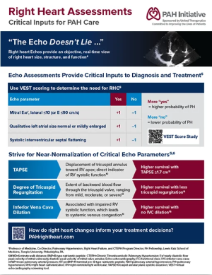
Right Heart Infographic
Use this infographic to review critical right heart assessments for PAH care.
Download Infographic
Right Heart Parameters Guide
Review this guide to learn more about critical echo and RHC parameters.
Download Guide
Watch an expert discuss the critical use of right heart echoes in clinical practice
Watch Short Video
Echo assessments provide critical inputs to diagnosis and treatment
Echoes in PAHThis content was developed based on ASE Guidelines.