Get the latest information with event videos and resources
Access the latest PAH Initiative event videos and resources right here! The PAH Initiative presents numerous educational events featuring nationally recognized experts in PAH care and treatment. Now, you can watch events anytime using the videos on this page. Each video covers a different topic to help keep you up to date on this complex disease. You’ll also find other helpful resources below, including quick summary sheets about each event.
Be the first to hear when new events are scheduled by signing up for PAH Initiative updates.
Event videos
Event summaries
Download the event summaries below for quick access to highlights from each event. If you have any questions about the topics covered, you can easily bring the summary sheet to your next appointment to discuss with your healthcare provider.
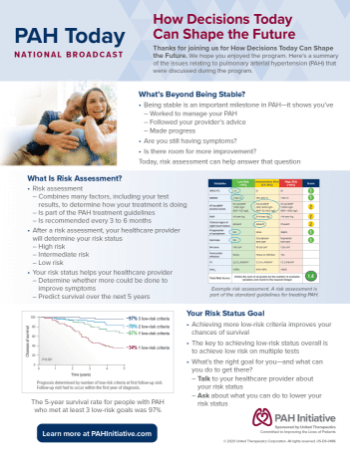
How Decisions Today Can Shape the Future
Learn why understanding your risk status is important for your PAH now and in the future.
Download Summary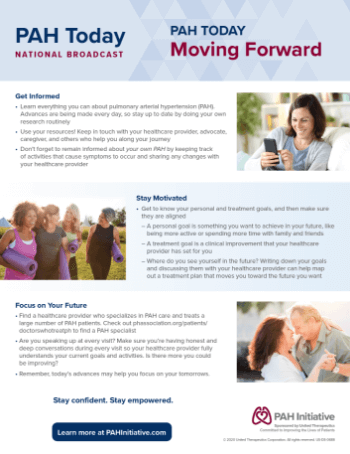
Moving Forward
Learn more about the steps you can take to stay informed and motivated to continue your treatment journey.
Download SummaryJoin the PAH Initiative
Sign up to stay up-to-date on events and all that the PAH Initiative has to offer.
Join the PAH Initiative